
Normally, the retina lines the inside of the eyeball, its nerve cells convert light into nerve impulses and send them to the brain along the optic nerve.
Retinal Detachment is a serious disease that requires urgent treatment. The main treatment for this pathology is surgery.
Retinal Detachment is a separation of the layer of photoreceptor cells – rods and cones – from the outermost layer – the retinal pigment epithelium, due to the accumulation of fluid between them.
This disrupts the nutrition of the outer layers of the retina, which leads to rapid loss of vision.
When the retina is detached, intraocular fluid enters under its layers, they cease to receive nutrition and die, which leads to blindness.
The risk of retinal detachment increases with myopia, the presence of retinal dystrophies, previous eye surgeries, eye injuries, diabetes mellitus, and vascular diseases.
Symptoms of Retinal Detachment:
Harbingers of retinal detachment can be: the sensation of light flashes in the eye, the curvature of straight lines. If the retinal vessel ruptures, then the patient may complain of the appearance of a large number of “flies before the eye”, black dots.
When a retinal detachment occurs directly, a dark shadow, curtain, veil appears before the eyes. Vision is rapidly deteriorating. In the morning hours, some patients note an improvement in visual acuity and an expansion of the field of view.
In a patient with a detached retina, a black veil appears that obscures part of the field of vision in the diseased eye, spreading to the entire retina, the eye completely stops seeing.
If there is a suspicion of retinal detachment, a comprehensive examination of the patient is required. Early diagnosis of retinal detachment helps prevent inevitable loss of vision.
At-risk groups:
• Patients with myopia and high astigmatism;
• Patients with established diagnosis of “peripheral retinal dystrophy”;
• Diabetes (diabetic retinal disease);
• Patients with neoplasms and hemorrhages;
• Heredity.
Treatment Methods:
Retinal Detachment is a disease that requires urgent treatment! With a long-term retinal detachment, persistent hypotension of the eyeball, cataracts, chronic iridocyclitis, subatrophy of the eyeball and incurable blindness develop. The main task in the treatment of detachment is the convergence of the layers of the retina. If there is a gap, it must be blocked.
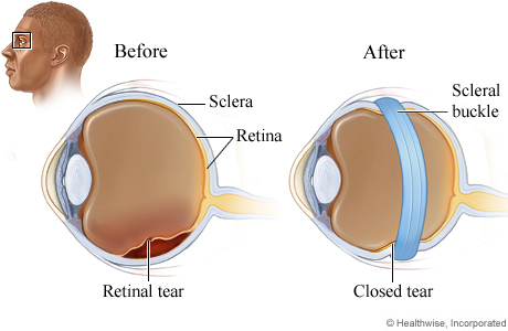
All methods of retinal detachment surgery are divided into external (scleral filling), when the intervention is performed on the surface of the sclera, and internal (vitrectomy), when the intervention is performed from the inside of the eyeball.
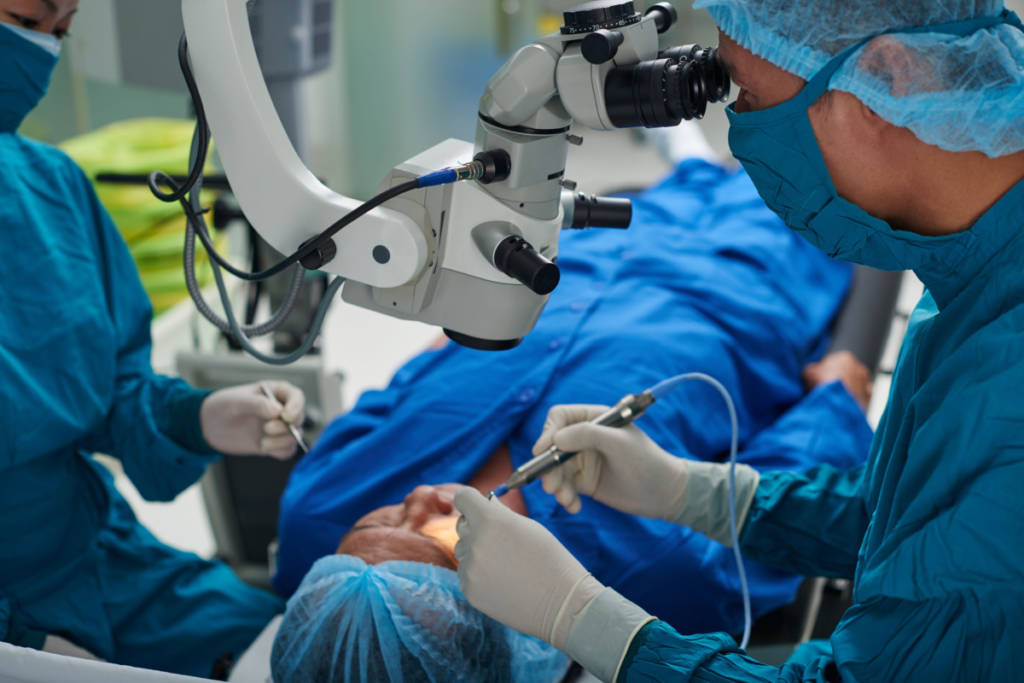
The most modern way to treat retinal detachment is vitrectomy – removal of the vitreous body with a temporary injection of silicone oil or gas into the eye cavity to ensure the retinal detachment fits.
Endovitreal surgery is an operation performed from the side of the eye cavity. When performing endovitreal intervention, access to the vitreous cavity and retina is provided through three scleral incisions less than 1 mm long, through which an illuminator, an instrument and a solution are introduced to maintain the tone of the eyeball.
When filling the sclera, the layers of the retina come closer together by creating an area of depression of the sclera from the outside. In the projection of the rupture of the retina, a silicone strip (filling) of the required size is attached to the sclera by means of sutures. In this case, the sclera under the strip is pressed inward, the sclera and choroid approach the retina, the created depression shaft blocks the gap, and the fluid accumulated under the retina gradually resolves.
Sometimes circlage is used – a circular impression with an elastic silicone thread or braid in the region of the equator of the eyeball. In some cases, with a large volume of accumulated subretinal fluid, it may be necessary to remove it (drainage) through a small puncture of the sclera.
The prognosis for vision depends on the age of the retinal detachment, the location of the breaks, and the state of the vitreous body. The optimal time for surgery is in the first few days and no more than 2 months in case of non-adhering retinal detachment as a result of a previous operation.
All detachments that have not fully adhered as a result of previous operations can and should be treated if no more than 1 year has passed since the detachment and the eye sees the light with confidence. If the eye does not see the light, then, as a rule, it is impossible to help.
After surgical treatment of retinal detachment, the magnitude of myopia and astigmatism often increases. Repeated retinal detachments (relapses) may occur. With relapses of retinal detachment, it is necessary to carry out repeated surgical operations, which are also not always effective.
The earlier competent surgical treatment is carried out, the better results it gives and the more it is possible to restore vision! The most favorable forecast for the restoration of vision after treatment is when the detachment did not have time to reach the central zone.
If the detachment managed to close the central zone, after a successful operation, the central vision, unfortunately, will not be able to fully recover.
Patients operated on for retinal detachment should be under the supervision of an ophthalmologist and avoid physical overload.
Prevention of retinal detachment:
The main preventive measure is a timely visit to an ophthalmologist when the first symptoms of retinal detachment appear and regular preventive examinations in the presence of risk factors.
After an eye injury, a complete ophthalmological examination should be performed. Examining pregnant women and performing prophylactic laser photocoagulation, if necessary, can also prevent retinal detachment during childbirth. Patients with high myopia, dystrophic changes in the retina or those operated on for retinal detachment are contraindicated in some sports, especially contact sports, as well as weight lifting.
Benefits of choosing AstraMedicaGroup
Only top qualified doctors
JCI certificated hospitals
Free COVID-19 Test before departure
4 nights in a 5-star hotel in Istanbul
Costs for laboratory, medication and equipment
Pre/post-operative tests
Free Istanbul tours
Latest technologies
Excelent travel assistance
All-round VIP transfer
Credit / Debit cards accepted
No prepayment
Personal assistants speak in English
24/7 customer service